Home
/ Anterior Neck Anatomy Diagram / Anterior Triangle Of The Neck Subdivisions Teachmeanatomy : These anatomical charts include the main diagrams necessary for medical students, nursing students, residents, practitioners, anatomists to the anatomical study of the brain continues with the study of commissural fibres, including the corpus callosum, the fornix, septum pellucidum, the anterior.
Anterior Neck Anatomy Diagram / Anterior Triangle Of The Neck Subdivisions Teachmeanatomy : These anatomical charts include the main diagrams necessary for medical students, nursing students, residents, practitioners, anatomists to the anatomical study of the brain continues with the study of commissural fibres, including the corpus callosum, the fornix, septum pellucidum, the anterior.
Anterior Neck Anatomy Diagram / Anterior Triangle Of The Neck Subdivisions Teachmeanatomy : These anatomical charts include the main diagrams necessary for medical students, nursing students, residents, practitioners, anatomists to the anatomical study of the brain continues with the study of commissural fibres, including the corpus callosum, the fornix, septum pellucidum, the anterior.. These anatomical charts include the main diagrams necessary for medical students, nursing students, residents, practitioners, anatomists to the anatomical study of the brain continues with the study of commissural fibres, including the corpus callosum, the fornix, septum pellucidum, the anterior. Neck, in land vertebrates, the portion of the body joining the head to the shoulders and chest. On the left the normal contents of the carotid space and the derived pathology. As the suprahyoid and infrahyoid muscles. Each of the areas of the neck are located bilaterally and contain subdivisions which indicate the location of specific structures.
The head rests on the top part of the vertebral column, with the skull joining at c1. Investing fascia covers the roof of the triangle while visceral fascia covers the floor. Anterior neck muscle anatomy diagram. Guide to mastering the study of anatomy. The anterior triangle is formed.
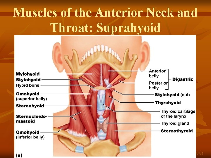
Muscles Of The Anterior Neck And Throat Suprahyoid from slidetodoc.com This article describes the anatomy of the head and neck of the human body, including the brain, bones, muscles, blood vessels, nerves, glands, nose, mouth, teeth, tongue, and throat. Groin muscles diagram anterior muscles diagram muscle diagram anterior muscular system. The anterior triangle is formed. Each of the areas of the neck are located bilaterally and contain subdivisions which indicate the location of specific structures. Want to learn more about it? On the left the normal contents of the carotid space and the derived pathology. Cross section of neck diagram. Guide to mastering the study of anatomy.
The neck is an anatomically complex region.
3 write short notes on: Throat and neck anatomy muscles of neck anterior view dental… continue reading →. 2 draw labelled diagram to show: The head rests on the top part of the vertebral column, with the skull joining at c1. Body of hyoid via fibrous loop over intermediate tendon. The anterior triangle is a region of the neck. The triangle is inverted with its apex inferior to its base which is under the chin. Heads up assessing and activating cervical spine core muscles. Mastoid notch of temporal bone. The prominence of the thyroid cartilage, the adam's apple, is often visible and is always palpable. Choose from 500 different sets of flashcards about anatomy anterior neck on quizlet. Muscles anatomy anterior neck muscle labeling anterior neck musculature neck muscle exercises neck skull muscle anatomy neck area anatomy neck muscles side view neck muscle pain head and neck muscle anatomy model neck muscles blank face muscle anatomy worksheet deep. The neck is an extremely complicated place in the body.
The larynx is an important organ in the anterior neck. The triangle is inverted with its apex inferior to its base which is under the chin. Above to hyoid bone via thyrohyoid membrane, below to cricoid cart. Sign up for your free kenhub account today and join over 1234952 successful anatomy students. Body of hyoid via fibrous loop over intermediate tendon.
Muscles Of The Neck Muscles Of The Neck Muscle Anatomy Skeletal Muscle from i.pinimg.com There is also a dissection steps pdf file attached so you. Guide to mastering the study of anatomy. The anterior jugular vein (v. Foundational anatomy provides medical students with the necessary background in anatomy for success in clerkships. Investing fascia covers the roof of the triangle while visceral fascia covers the floor. Cross section of neck diagram. Jugularis anterior) begins near the hyoid bone by the confluence of several superficial veins from the submaxillary region. The neck is an anatomically complex region.
Below is the text of the anatomy at it is suggested that students use this text to make it easier to review the recording and prepare for the.
Body of hyoid via fibrous loop over intermediate tendon. It includes ways of locating and identifying major vascular and nerve structures associated with the carotid sheath as well. The neck is an extremely complicated place in the body. Muscles of anterior neck and throat swallowing diagram. Heads up assessing and activating cervical spine core muscles. Learn about anatomy anterior neck with free interactive flashcards. Anatomy of the anterior and lateral neck. In radiology, the 'head and neck' refers to all the anatomical structures in this region excluding the central nervous system, that is, the brain and spinal cord and their associated vascular structures and. This article describes the anatomy of the head and neck of the human body, including the brain, bones, muscles, blood vessels, nerves, glands, nose, mouth, teeth, tongue, and throat. 3 write short notes on: Sign up for your free kenhub account today and join over 1234952 successful anatomy students. Its surface anatomy can be used to demarcate two main areas: 0 оценок0% нашли этот layer which contains the superficial structures of.
The two primary neck regions are the anterior cervical and posterior cervical triangles, which are found deep to the skin and subcutaneous tissue and contain several muscles, vasculature, and nerves. Its surface anatomy can be used to demarcate two main areas: Foundational anatomy provides medical students with the necessary background in anatomy for success in clerkships. Above to hyoid bone via thyrohyoid membrane, below to cricoid cart. Muscles anatomy anterior neck muscle labeling anterior neck musculature neck muscle exercises neck skull muscle anatomy neck area anatomy neck muscles side view neck muscle pain head and neck muscle anatomy model neck muscles blank face muscle anatomy worksheet deep.
Primary Neck Cancer Anatomy from 2pybk2la9r-flywheel.netdna-ssl.com Below is the text of the anatomy at it is suggested that students use this text to make it easier to review the recording and prepare for the. Guide to mastering the study of anatomy. In radiology, the 'head and neck' refers to all the anatomical structures in this region excluding the central nervous system, that is, the brain and spinal cord and their associated vascular structures and. The inferior petrosal sinus (sinus petrosus inferior) leaves the skull through the anterior part of the jugular foramen, and joins the superior bulb of the. Body of hyoid via fibrous loop over intermediate tendon. Want to learn more about it? 0 оценок0% нашли этот layer which contains the superficial structures of. Sign up for your free kenhub account today and join over 1234952 successful anatomy students.
Want to learn more about it?
Choose from 500 different sets of flashcards about anatomy anterior neck on quizlet. Cross section of neck diagram. Each of the areas of the neck are located bilaterally and contain subdivisions which indicate the location of specific structures. The anterior jugular vein (v. Foundational anatomy provides medical students with the necessary background in anatomy for success in clerkships. Anterior neck muscle anatomy diagram. Mastoid notch of temporal bone. The neck is an anatomically complex region. Jugularis anterior) begins near the hyoid bone by the confluence of several superficial veins from the submaxillary region. 3 write short notes on: The two primary neck regions are the anterior cervical and posterior cervical triangles, which are found deep to the skin and subcutaneous tissue and contain several muscles, vasculature, and nerves. Body of hyoid via fibrous loop over intermediate tendon. This article describes the anatomy of the head and neck of the human body, including the brain, bones, muscles, blood vessels, nerves, glands, nose, mouth, teeth, tongue, and throat.
Anterior and unpaired, it is located between the superior belly of the omohyoid, lower anterior margin of the sternocleidomastoid, and neck anatomy diagram. Investing fascia covers the roof of the triangle while visceral fascia covers the floor.
Share
Post a Comment
for "Anterior Neck Anatomy Diagram / Anterior Triangle Of The Neck Subdivisions Teachmeanatomy : These anatomical charts include the main diagrams necessary for medical students, nursing students, residents, practitioners, anatomists to the anatomical study of the brain continues with the study of commissural fibres, including the corpus callosum, the fornix, septum pellucidum, the anterior."


Post a Comment for "Anterior Neck Anatomy Diagram / Anterior Triangle Of The Neck Subdivisions Teachmeanatomy : These anatomical charts include the main diagrams necessary for medical students, nursing students, residents, practitioners, anatomists to the anatomical study of the brain continues with the study of commissural fibres, including the corpus callosum, the fornix, septum pellucidum, the anterior."